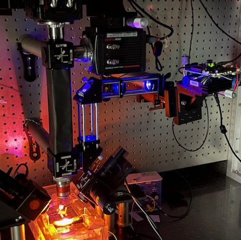
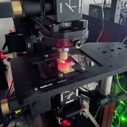
Our custom made random access non-linear microscope allows operation in both two-photon fluorescence (TPF) and second-harmonic generation (SHG) imaging modes. It is equipped with a tunable femtosecond laser and an advanced scanning system for high-speed, multi-region imaging.
Main features:
• Rapid Multi-Region (RMR) scanner
• Backward and forward detection
• Polarization anisotropy measurements
• Three-channel acquisition
Our mesoscale selective plane illumination microscopy (mesoSPIM) is composed of two custom-designed telecentric lenses and two high-performance back-illuminated sCMOS sensors for double-side light collection, resulting in an object field of view of approximately 1 cm² - sufficient to acquire an entire longitudinal cross-section of cleared murine hearts with negligible field -curvature distortion and a resolution of 3.25 um.
Main features:
• Dual-side illumination and detection
• Four excitation wavelengths
• Complete reconstruction of entire cleared hearts in a single-scan
• Compatible with SHIELD and CUBIC tissue-clearing protocols
Our Zeiss Axio Zoom.V16 system, equipped with an Apotome structured illumination module, enables high-resolution fluorescence imaging across large fields of view. The system combines zoom optics with optical sectioning capabilities, making it ideal for thick tissue samples and cleared organs.
Main features:
• High-resolution zoom microscopy with large depth of field
• Structured illumination for optical sectioning (Apotome)
• Suitable for imaging thick sections and cleared tissues
• Fluorescence imaging across multiple channels
A dedicated facility optimized for processing cardiac tissue for high-resolution 3D imaging. The system enables whole-organ transparency and efficient antibody penetration using advanced clearing protocols, while preserving structural and molecular features.
Main features:
• Optimized for cardiac tissue architecture
• Compatible with CLARITY, SHIELD, and CUBIC clearing protocols
• Enables deep antibody labeling for immunostaining
This custom-built system enables simultaneous monitoring of transmembrane voltage and cytosolic calcium, while performing optogenetic stimulation in whole-heart preparations. Designed to minimize spectral crosstalk, it allows precise functional interrogation of cardiac electrophysiology using an all-optical approach.
Main features:
• Dual imaging of voltage (ElectroFluor 730p) and calcium (X-Rhod-1)
• Integrated optogenetic control via Channelrhodopsin-2 (ChR2)
• Minimal interference using the isosbestic point of ElectroFluor 730p
Custom-built all-optical platform designed for panoramic optogenetic stimulation and simultaneous voltage imaging across the entire surface of the heart. The system integrates four DMD-based projectors and four high-speed sCMOS cameras, enabling precise and spatially continuous stimulation without mechanical contact.
Main features:
• 360° stimulation and imaging using four DMDs (TI LightCrafter 4500) and four sCMOS cameras (Hamamatsu ORCA-Flash4.0 V3)
• High temporal (≥1 kHz) and spatial (~80 μm) resolution
Best AI Website Maker